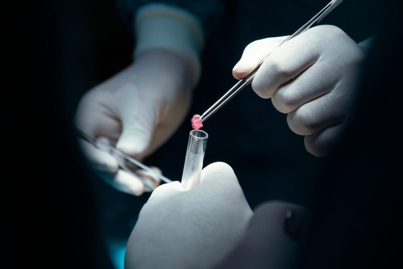
Renal or kidney biopsy is a medical procedure involving taking a small bit/tissue sample from the kidney for diagnostic purposes. Typically performed under imaging guidance, such as ultrasound, under local anesthesia, kidney biopsy helps identify and analyze various disorders, including glomerular diseases and renal inflammation. This procedure aids in determining the underlying cause of kidney dysfunction, guiding appropriate treatment decisions.
Renal biopsy plays an important role in nephrology, allowing clinicians to tailor interventions, monitor disease progression, and optimize patient care based on precise pathological information obtained from the sampled kidney tissue.
Types of Renal Biopsies

Renal biopsies are essential diagnostic procedures for kidney diseases.
Two primary types of renal biopsies are:
Percutaneous Kidney Biopsy
Percutaneous kidney biopsies, performed under local anaesthesia, involve inserting a needle through the skin into the kidney to obtain a tissue sample. This method, often guided by imaging techniques like ultrasound or CT scans, is less invasive, reducing recovery time.
Open biopsy (surgical biopsy)
Open surgical biopsies are rarely done and involve a small surgical incision to directly access the kidney. This method may be preferred in certain situations, such as when a percutaneous biopsy is challenging due to anatomical factors.
While percutaneous biopsies are generally more prevalent due to their minimally invasive nature, the choice between the two depends on the specific clinical scenario and the preferences of the medical team.
Objective of Renal Biopsy Diagnostic procedure
The primary purpose of a renal biopsy is to obtain detailed information about the structure and function of the kidneys. It is often recommended when there is suspicion of kidney disease based on symptoms, laboratory tests, or imaging studies.
Common indications for a renal biopsy include unexplained proteinuria (excessive protein in the urine), hematuria (presence of blood in the urine), rapidly declining kidney function, or the presence of abnormal findings in imaging studies such as ultrasound or CT scans.
Through histological analysis of the biopsy specimen, healthcare professionals can differentiate between different types of glomerulonephritis, assess the extent of inflammation, and identify any underlying causes such as autoimmune disorders or infections. This information is necessary for tailoring appropriate treatment strategies, guiding prognosis, and determining the overall management of kidney diseases.
Furthermore, renal biopsy aids in distinguishing reversible from irreversible kidney damage, aiding in therapeutic decisions. It plays an important role in evaluating the effectiveness of interventions and adjusting treatment plans accordingly.
Overall, the purpose of a renal biopsy is to provide precise diagnostic information, enabling healthcare providers to make informed decisions and optimize care for patients with kidney disorders.
How does one prepare for Renal Biopsy?
Before undergoing a renal biopsy, patients typically undergo a series of preparations to ensure a safe and effective procedure. The following are the various aspects to be considered:
- Medical evaluation: A thorough medical history is obtained to identify any pre-existing conditions, allergies, or medications that might affect the procedure. Blood tests may be conducted to assess clotting function and kidney function.
- Informed consent: Patients are informed about the procedure, its purpose, potential risks, and benefits. They must provide informed consent before the biopsy.
- Fasting: In most cases, patients are not required to fast before the biopsy. However, they are supposed to take a light breakfast before the procedure, as the procedure entails them to lie on their stomach
- Medication adjustment: Certain medications, especially those affecting blood clotting, may need to be adjusted or temporarily discontinued before the biopsy. This is done under the guidance of healthcare providers.
- Imaging studies: Imaging studies, such as ultrasound or CT scans, may be performed to visualize the kidneys and guide the biopsy needle accurately to the target area.
- Blood pressure monitoring: Ensuring blood pressure is well-controlled is important, as uncontrolled hypertension can increase the risk of bleeding during and after the biopsy.
- Emptying bladder: Patients are often instructed to empty their bladder before the procedure to facilitate access to the kidney.
- Transportation arrangements: Since patients may experience mild discomfort or pain after the biopsy, arranging for transportation home is advisable.
Individuals must communicate openly with their healthcare team, providing accurate information about their health and following pre-biopsy instructions carefully. These steps contribute to a safer and more effective renal biopsy process.
What happens during the Renal Biopsy process?
The following are the various steps involved in a renal biopsy:
- Patient preparation: Blood tests are often conducted to check clotting factors and assess the overall health of the patient.
- Consent: The patient is informed about the procedure, its purpose, potential risks, and benefits. Informed consent is obtained before proceeding.
- Positioning: The patient is positioned, often lying on their stomach or side to expose the lower back area.
- Anaesthesia: Local anaesthesia is administered to numb the skin and underlying tissue at the biopsy site. In some cases, mild sedation may also be given to help the patient relax.
- Imaging guidance: Using imaging techniques like ultrasound or fluoroscopy, the healthcare provider locates the kidneys and identifies an optimal entry point for the biopsy needle.
- Biopsy needle insertion: A thin biopsy needle is carefully inserted through the skin and into the kidney to collect a small tissue sample. This process may be guided by real-time imaging to ensure accurate placement.
- Tissue collection: The needle is quickly and carefully advanced into the kidney to retrieve a small core of tissue. This tissue sample is then sent to the laboratory for microscopic analysis.
- Post-biopsy observation: After the procedure, the patient is observed for a few hours to monitor for any signs of complications, such as bleeding or pain.
Recovery after a Renal Biopsy
After a renal biopsy, patients typically experience a short recovery period.
Immediately post-procedure, patients are observed for a few hours to monitor for potential complications such as bleeding. Mild discomfort or pain at the biopsy site is common and can be managed with prescribed pain relievers.
Rest and avoiding strenuous activities for a day or two are advised to minimize the risk of bleeding. Follow-up appointments are scheduled to discuss biopsy results and determine appropriate treatment plans based on the findings.
Patients must adhere to post-biopsy care instructions provided by healthcare providers for a smooth recovery and optimal healing of the biopsy site.
Risks of a Renal Biopsy
Renal biopsies, while generally safe, do carry certain risks and potential complications. The primary concern is bleeding, ranging from minor to severe cases requiring intervention.
Pain, infection, arteriovenous fistula formation, and pneumothorax (rarely) are also possible complications. Hematoma formation and transient hematuria (blood in urine) may occur. Allergic reactions to medications used during the procedure are rare.
Although these risks are generally low, careful consideration and informed consent are important before undergoing a renal biopsy. Healthcare providers employ sterile techniques and close monitoring to minimize and manage potential complications. Consult your healthcare provider immediately, if you are having:
- Bright red blood in your urine for longer than 24 hours after biopsy
- Difficulty in urinating
- Chills or a fever
- Pain at the biopsy site that increases in intensity
- Redness, swelling, bleeding, or any other discharge from the biopsy site
- Weakness
Conclusion
Renal biopsy results provide valuable insights into kidney health, aiding in the diagnosis and management of various renal conditions. Pathologists examine the tissue sample under a microscope, assessing cellular structures to identify abnormalities. If the renal biopsy sample shows a normal kidney structure that is free from abnormal deposits, the results are considered normal. If the renal biopsy sample shows changes in the kidney tissue, it is considered abnormal. If the results are abnormal, it could be a sign of:
- Infection of the kidney
- Decreased flow of blood to the kidneys
- Connective tissue disorders
- Failure of a kidney transplant
- Urinary tract Infection (complicated)
- Kidney cancer
- Disease conditions that affect kidney function
Results guide tailored treatment plans, including medications, lifestyle modifications, or further interventions. Effective communication between healthcare providers and patients is important for understanding the implications of biopsy results and making informed decisions about managing kidney health.
Regular follow-up assessments, monitor the progression of renal conditions and adjust treatment strategies as needed.
NU Hospitals in Bangalore, India, is renowned for excellence in kidney care. With a specialized focus on nephrology and urology, NU Hospitals stands out for its expert medical professionals, cutting-edge technology, and patient-centric approach.
The hospital's commitment to delivering top-notch renal services has earned it a reputation for excellence in kidney care. Patients seeking quality renal biopsy procedures and comprehensive kidney healthcare often turn to NU Hospitals for their expertise and exceptional medical services.
Renal biopsies are essential diagnostic procedures for kidney diseases. Two primary types of renal biopsies are:
Author: Dr. Nikhil J Elenjickal

